Posts

Showing posts from June, 2017

Week 2: MRI and Orthopedics Research

Image
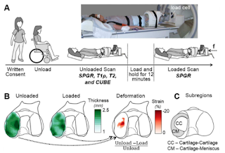
This week I had the opportunity to observe some MRI scans for various research projects. On Monday we performed a unique scan of a patient who had previously received a meniscal graft. The patient's knee joint was loaded to half their body weight using an MRI compatible force plate and imaged to detect localized compression of the tissue. These strain maps may eventually be used to determine whether the graft is effectively distributing loads or if a revision may be necessary. In addition to imaging patients, the MRI lab scans animal tissues which often come from the Cornell University vet school. This often requires special preparation to enable the use of certain coils and protect both the scanner an sample. Last week were able to scan a horse hoof using ultrashort echo time (UTE) imaging techniques (see Week 1 blog post). This week I studied different methods to quantify T2 relaxation times in tendon using these images. On Thursday we attempted to scan rat patellar tendons tha

Week 2: The OR - A Memoir from a Biomedical Engineer

Image
The second week of the Immersion Program was all about discovery! In less than five days, I was able to briefly cover the entire spectrum of a hip arthroplasty, a surgical reconstruction or replacement of a joint. It all started on Tuesday by learning about the use of patient-specific triflange designs in cases where patients have a surgical revision after a total hip arthroplasty (THA). Patients may need to have a THA revision due to post-surgical complications such as instability, aseptic loosening, osteolysis and/or wear, and infection (Springer et al., 2009). A patient-specific triflange is often considered as an alternative solution when a severe bone loss prevents the reconstruction of the joint using other well-established surgical techniques. The term triflange is used to describe the three areas where the implant is attached to the hip bone: ilium, ischium, and pubis (Fig. 1). During the presentation hosted by the Biomechanics Lab Meeting, I learned that design engineering is

Week 2

Image
First of all, some updates on my project, 2-photon zebra fish neural image segmentation. As I have mentioned in last week’s blogger, I have tried a couple simple algorithms, like simple thresholding and watershed. Here is the results of them: Fig 1: Thresholding results.  Thresholding (Fig 1). This algorithm is easy to understand, and is usually used as a preprocessing step. Watershed. It is a commonly used segmentation algorithm. Intensity of image is considered to be height in the topography. As water is flooded into the topography, different catchment basins are separated when different flooded regions started to merge. Here, this algorithm is implemented in two different ways: Thresholding; generating the distance map based on the binary image; applying watershed to the resulting distance map. (Fig 2) Fig 2: Distance map (left) and watershed segmenting results (right). Fig 3: Image imposed with regional minima (left) and watershed segmenting results. 

Week Two: Tracking Tops

Image
               Last week I wrote about MRI/US Fusion Guided Biopsy, but one thing I did not go into detail was the equipment used during the biopsy. In general, the equipment involved are the biopsy needle, the ultrasound probe, and the fusion biopsy device itself. The first two pieces of equipment are well known, but the fusion biopsy device is not always recognizable. A fusion biopsy device, in general terms, is the centerpiece of the procedure. Overall, it is responsible for tracking the position of the ultrasound probe, displaying the segmented MRI and US images, and recording the location of the biopsies. All these functions make it more impressive the number of different approaches companies have employed to execute these demands, specifically in tracking the US probe position. Currently, there are three methods to achieve this: i) mechanical systems, ii) electromagnetic tracking, and iii) image-based tracking (examples shown below). Here, I have observed biopsies with mechanica

Week 2: They have neat toys in NYC

Image
The Nikon Biostation:  An automated cell culture imaging system that can take images of cell culture plates at scheduled time periods in both brightfield and in fluorescent spectra. Images taken from product website.  This week I got to live a life of luxury for a tissue engineer. I started out my fabricating organoids for the primary human umbilical vein endothelial cells and the immortalized lymphatic endothelial cells that I have been growing in 2D monocultures for the last week. They turned out great and my lab was really grateful to have someone specializing in 3D tissue cultures to add a robustness to their research that they didn't have the skills to do on their own. If these cells were grown in the right conditions, they have the potential to form vessel-like structures through self organization. Normally at home in Ithaca, that would mean I would check my organoids multiple times a day, taking pictures of every well every time. In a 96-well plate full of samples this

Week 2: Surgical Pathology

Image
Over the last two weeks, I have been trying to build a surface-level understanding of pathology and the techniques used to diagnose. One of my professors in undergrad would always tell us that in order to really understand a technique, we have to understand how and why it came to be. So this week, I read a little bit about the history of pathology. Here’s what I found out: Pathology started as simply an observation of the body. Early physicians would keep track of symptoms and relate them to what they saw on (or in) the patient’s body. They could get a rough idea of the causes and effects of a dise ase.  This was the easiest way for them to get the information they wanted. The microscope is extremely prevalent in this field, and this invention allowed the study of pathology to expand to a cellular (and with newer technology, atomic) level. In the late 19 th century, scientists were just beginning to understand how biology worked on this small scale, and,  through many differe

Week 2

My second week of being immersed involved meeting with patients during Dr. Spector’s clinic days and observing more surgeries. One interesting case involved a patient with a large tumor on their neck. The tumor had to be excised and the resulting void in tissue must be replaced. After the head and neck surgeon carefully removed the tumor and nearby lymph nodes, Dr. Spector’s team worked on repairing the resulting defect. This was accomplished by using a technique known as a rotated flap. During the procedure, Dr. Spector mobilized the patient’s pectoral muscle and moved it to the neck where the tumor was previously located. However, the muscle wasn’t fully detached to keep a blood supply to the rotated flap. This would allow the muscle to integrate into the new wound and facilitate repair by introducing a healthy blood supply. This procedure contrasts with a free flap, where tissue can be completely removed from distant parts of the body and placed on the wound. In this case, the surge

Week 2: First time to watch surgeries

Image
Fortunately, the issue with an account for scrubs was cleared quickly, so I was able to observe two surgeries. http://www.houstonmethodist.org/orthopedics/where-does-it-hurt/ankle/ankle-fusion/ The first surgery was "ankle arthrodesis," also known as ankle fusion, which is to remove articular cartilage between bones, fibula, tibia, and talus and combine together. The ankle arthrodesis is usually performed in order to alleviate pain and maintain or improve the movement of a patient. In the case of severe ankle arthritis, this surgery is recommended since it takes long time to get recovered and could limit some activities.There were total three clinicians specialized in each step for this surgery. The first impression of the OR was that the atmosphere was natural. For example, they played songs, and sometime they made some jokes to make themselves relax, not too tense. Of course, they were serious during the surgery as well. http://orthoinfo.aaos.org/topic.cfm?to

NYC Chronicles (Week 2): Medicine is a Team Sport

This week at Columbia University Medical Center (CUMC), I  joined Dr. Prince at his weekly meeting with Dr. Graham Barr, an associate medical professor and a specialist in respiratory epidemiology, and other collaborating members to discuss the use helium-3 diffusion weighted imaging ( 3 He-DWI) to calculate the size of alveoli in patients with emphysema. As I mentioned before in my previous post, DWI is a form of MR imaging that relies on the Brownian motion of protons in tissues to obtain images, displaying intensified regions of restricted proton diffusion, which typically occurs in pathological areas. 3 He-DWI can be used as an alternative or a supplement to traditional MR imaging to scan regions of the body with low proton density, such as the lungs, to further characterize the healthy and diseased tissues of patients. By attending this meeting, I noticed that there were subtle differences in problem-solving and critical thinking, as well as diverse preferences in experimental de

Week 2: Settled in the City

Image
Tibra Wheeler Two weeks down, five more to go – my time in the city is flying by way too fast. Before I talk about this past week, let’s talk about my first weekend in NYC was definitely an experience. We traveled to Brooklyn to go to Smorgasburg, which is an outdoor foodie market on Saturdays. There were so many food options in one area that it was overwhelming (I’m not one for making decisions when it comes to food). I finally settled on getting chicken and waffles. While eating that deliciousness, I waited in line (for a long time) for John’s Juice to get a Pineapple John. It was some of the best pineapple juice I’ve ever had plus it was served in the pineapple with a super cute straw. We walked around Brooklyn, getting some great views of the city from the East Shoreline and took a tour of the Boston Brewery to learn some history. That evening, we went to Vinnie’s pizzeria and I got a pizza that had mini pizzas as the toppings – “pizza on top on pizza.” I took a Snapchat pictu

Week 2: Cut short by illness :(

My second week in New York began with observing a lot of interesting work, and took an unfortunate turn as I came down with a bit of illness around Wednesday afternoon. On Monday, I had the opportunity to meet with Dr. Vivek Mittal’s group and discuss how they were working in parallel with Dr. Vahdat’s clinical trials to study the effects of copper depletion on the pre-metastatic niche in breast cancer.  This was complemented by my shadowing of Dr. Vahdat on Wednesday where we met with a number of patients who were still in progress as part of this trial.  Alongside Dr Vahdat, I have found it very interesting how decisions are made regarding the selection of treatment options over the course of cancer therapy.  Similar to my experiences last week, I was consistently impressed by how well the patients had researched their treatment options and were able to discuss the crucial factors of treatments, such as side effects and duration of treatment, that I was not keenly familiar with.

Week 2: Let the Immersing Continue

Image
Since the end of last week, I have continued to observe rounds in the cardiology ICU during the mornings. These cases continue to be very interesting as many patients come in with a variety of complications and the doctors have to determine the best course of treatment including the drugs they will be administered and what specific diagnostic procedures are necessary to determine the underlying causes of their symptoms. I am really enjoying being a "fly on the wall" and listening in on the conversations the doctors have together as they attempt to diagnose and treat these patients as soon as possible. Besides continuing to observe rounds, I have also set up my research project this summer. I met with Dr. Gaudino and his fellows, Dr. Ohmes and Dr. Leonard. As part of my clinical research, I will be performing a meta-analysis on patients who have reoccurring cardiac surgeries after repair of their ascending aortic aneurysm and/or dissections. An aortic aneurysm is essentially

Second Week of Immersion

Image
For this week, my clinical mentor, Dr. Susan Gauthier, was out of town. In the meantime, I was working on creating the PET kinetic model with phantom data until I was able to receive actual data to begin analysis on. Outside of working on the project, I was able to shadow Dr. Ted Schwartz to see a neurosurgery being done where a tumor was removed from near the brain through a hole drilled along the wall of the patient's nose. What impressed me most of the surgery was not the procedure itself but one of the guiding tools used in which the patient's MRI was mapped to a probe that allowed the surgeons to accurately locate the tumor for removal. From what I had seen, the probe used electrode sites located on the head of the patient to calibrate the positioning of the probe to a previous MRI on file. From there, the surgeons could see on a screen, the MRI indicating the location of the tip of the probe, shown in below (Ivanov et al, 2017). Today, though, I was able to observe

Week Two (Jason Chang)

Image
This week, I focused on installing MATLAB onto my work computer and getting clearance to observe OR procedures at WCMC and the Hospital for Special Surgery (HSS). As far as clinical exposure this week, Carla , one of the stroke research coordinators, b rought me with her to the emergency room on Monday after she was paged about an incoming stroke patient. However, the patient turned out to be a construction worker who had fallen and sustained several blunt force injuries, which meant that the patient fell under the jurisdiction of trauma, not neurology. I t was very interesting to see the high-pressure and fast-paced environment that the emergency medicine clinicians work in. Additionally, Dr. Prince gave a lecture on Monday on interpreting imaging results, specifically with magnetic resonance imaging (MRI), computed tomography (CT), and x-ray scans. During his lecture, Dr. Prince emphasized the importance of always having multiple views for every scan to confirm any abnormalities

Carolyn: Lab work and more dessert!

Image
During my second week, I made progress in the lab as my clinician was traveling. I started to perfect my RNAseq techniques. Although I have several different protocols to use for reference, I am still working to ensure that the RNA I extract is of high quality. This remains a work in progress! I was able to attend several interesting meetings this week. At the Hospital for Special Surgery this morning, a physician from California gave a talk regarding a non-profit organization he runs, Global Spine Outreach. This organization travels to underprivileged areas and helps to provide expert spine surgeries to pediatric patients suffering from severe scoliosis. The images he showed were incredibly moving, as it is clear that these patients have almost no hope of normal life with their current standard of care. This surgeon makes a huge impact and changes their lives for the better. Additionally, he provides some instruction to the practicing physicians living in these areas, encouraging b

Week 2 - Moni

Image
Week 2 was more productive, since I've started getting clearances to do more lab work. Notably, on Tuesday, the lab spent the day doing mouse surgery to test the efficacy of a new cell line in healing tendon rupture of the rotator cuff. It took our 6 person team 12 hours since we did 10 mouse cages. I was impressed with the tenacity of the scientists in our lab to do this procedure on these incredibly small mouse samples through the entire day. I also got trained on the multiphoton microscope at WCMC, which I will be using in the coming weeks to evaluate tendon degeneration in mouse models. Today I was able obtain surgical discards of tendon samples from Dr. Rodeo's surgeries that I will be using for my studies to investigate mechanobiology of degenerative tissue. I also had the opportunity to meet some engineers from GE, who were designing new MRI coils for patient comfort. It was interesting for me to talk to the engineers and the radiologists to see common problems fac