Posts

Showing posts from July, 2017

Week 6: Un Cubano en Nueva York

Josue Santana It is hard to believe that we are almost at the end of the Summer Clinical Immersion Program. It feels like yesterday where I was running all over the hospital to get the security clearance to do my clinical project, access the hospital, and watch surgical procedures. Throughout this week, I worked primarily in my retrospective clinical research study. We finally completed the portion of the study where we searched for multiple demographic and skeletal-specific factor on the patient’s medical history. Our preliminary results look quite promising, and I am thrilled to run the statistical analysis of our data on the first days of the upcoming week. From our patient’s cohort, the biggest reason for a surgical revision after a total knee replacement was infection followed by instability, aseptic loosening, stiffness, and periprosthetic fracture. This preliminary finding is quite outstanding because it reinforces the importance of having rigorous protocols not only within t
Week 6

Our time in New York City approaches its end at the close of our 6 th week of being immersed. Last week, I talked about an alternative strategy to tissue grafting and flaps. The use of tissue grafts and flaps to fill defects in existing tissue is a tool that is widely employed by reconstructive and plastic surgeons. One surgery I witnessed this week demonstrated a prime example. In this case, the patient developed a tumor on their tongue that had to be excised. As with any typical head and neck procedure, this surgery would be relatively complicated with large of amounts of tissue either being removed or mobilized to completely remove the tumor. After the tumor was removed, the patient was missing most of their tongue, along with a large void inside their mouth and through their neck. To reconstruct the missing tissue, Dr. Spector mobilized a pectoral flap and rotated it into the position of the tissue defect, similar to a case I observed earlier in the summer. Additionally, part of

Week 6: Alex

Hello friends! This week was particularly interesting as I spent some time in the Clinical Microbiology laboratory within the Laboratory Medicine division of the hospital with Dr. Lars Westblade. Throughout the week, I was able to spend time at different workstations, learning how pathogens from different sources (wounds, blood, urine, etc.) grow on different culture mediums and how these growth patterns can help diagnose an infection. I also learned how antibiotic susceptibility testing is done and how it can help physicians properly treat patients. I was absolutely amazed at the diagnostic equipment they have. I remember in high school we would grow bacteria on an agar plate with different antibiotics to illustrate resistance patterns. Although that's still done here, what amazed me was the automated equipment they use here for diagnosis of infectious diseases. I got to see a Biofire in action, which is essentially a machine that can detect antibiotic resistance directly from c

Week 6

With only one week remaining in the city, I am beginning to feel the rush to do everything at once but that is not all a bad thing.   In the clinic this week, I had the opportunity to shadow several doctors in the radiation oncology ward where I got a complete overview of how radiation treatment is prepared, designed, and delivered to patients in order to precisely target tumor regions of interest.  Beyond the minutiae of preparations, I was impressed by how sophisticated the radiation machines have become in order to deliver precise doses of radiation to very specific geometrical regions of the body. Regarding my project, I have continued to organize the data and develop a matlab script which will be able to evaluate the impact of copper depletion on blood serum markers which are representative of remodeling within the tumor microenvironment.  By observing changes in the blood levels of lysyl oxidase, collagen-breakdown and collagen formation markers (such as matrix metalloproteina

Week 6 (Moni)

Image
This week was crunch time in lab, since I needed to start wrapping up my projects before leaving. For the multiphoton stage redesign, I took measurements and have acrylic sheets that I will use to cut and superglue together to give the greater range of motion to fit an animal in the imaging space. I also ran my last batch of flow cytometry today on the human surgical discards, and learnt how to use FlowJo to analyze my data. Next week I will get some statistics on this data to present for my immersion poster. On Wednesday, our lab did sheep knee dissections for a collaboration between our lab, Dr. Hollis Potter (HSS), and Dr. Lisa Fortier (Cornell Ithaca). These sheep underwent a menisectomy and the study is investigating the efficacy of a scaffold with various cytokines to aid in meniscus regeneration. After spending the summer watching surgeons operate, it was nice to finally do some surgery myself :) Intact sheep knee It was also a very exciting week for me to be out

Week 6: Pipetted in the City

Image
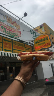
Tibra Wheeler Flashbacking to the 90’s this past weekend was really fun but back to the 21 st century for this girl this week. Over the weekend, we also went to Coney Island. We ate “The Original” Nathan’s Hotdogs, rode the 90-year old wooden roller coaster the Cyclone, visited the New York Aquarium, and walked along the boardwalk. The end of immersion is quickly approaching so this week was time to really focus on what goals I wanted to reach by the end of next week. It also means trying to fit in every tourist thing, every restaurant, and every dessert I want to do before I leave NYC. Monday was a day for me to do some more touristy things with some friends. We decided to take the Statue Cruises to Liberty Island and Ellis Island. I was really excited to go see Lady Liberty as well as visit the Ellis Island Immigration Museum. Unfortunately, tickets were sold out to go inside the Statue of Liberty, but it was still an experience. I’m also pretty sure we picked the win

Week Six: Soccer Sheets

Image
Another week of being immersed has come to an end and so has the week of coding. Now last week issues arose through differences in version numbers, but this week has been a different kind of coding. While I have my functions running gather data from different files, I have been coding in the sense that I have been assigning different values for different events seen in pathology reports. As that sentence suggests, this week has been a lot of looking over pathology reports to get out data regarding biopsy results. This has led to many hours spent on Excel putting in parameters related to prostate biopsies such as the Gleason scores (the pathological score given regarding the degree of differentiation related to cancer in prostate tissue). As a result, I have looked at several pathology reports to build the database related to my research. Fortunately, I have had some help with the rising second year medical students that have been working on this project with me. Without them this woul

Week 6

This week I worked on analyzing the MR images of rat tendon we were able to take last week. A unique challenge with these images was segmentation using ITK-SNAP. Whereas the scans of horse and pig tendon had taken UTE slices in the sagittal direction, we had to take slices in the axial direction for the rats, as this was the only direction in which multiple slices of the tendon could be taken in such a small animal. This made the cross sectional area of the tendon much smaller and difficult to differentiate in the image. In order to improve segmentation accuracy, I attempted to use a different pulse sequence with higher resolution to guide segmentation of the lower resolution UTE images. This required me to use an algorithm to "reslice" the higher resolution images to match the UTE images. Next week I hope to use these images to segment and finally quantify T2* values in the rat tendon. In addition, on Monday I had the opportunity to see multiple knee surgeries including a

Week 6

Image
This week, I also shadowed Dr. John Kennedy for the surgery and clinic as well. In terms of clinics, there were not many things different from what I have seen for last couple of weeks. Most therapies were shockwave or PRP injection. However, he explained more about the mechanisms of injuries, which was really helpful for me to understand how the treatment can be determined. Another thing I was impressed by was the Lipogems. http://www.jockdoctors.com/services/lipogems-lipoaspiration-treatment/  Bianchi, Francesca, et al. "A new nonenzymatic method and device to obtain a fat tissue derivative highly enriched in pericyte-like elements by mild mechanical forces from human lipoaspirates."  Cell transplantation  22.11 (2013): 2063-2077. This procedure using the Lipogems has gained a lot of attention due to its powerful therapeutic effect. Simply, first thing to do is aspirating adipose tissues from a patient, and purifying the essential components which are mainly mese

Week 6: Brains & Chocolate

Image
Just when I think I’m starting to get some of the basics down, I realize that there’s so much more to learn (as always). At first, four years of medical school and a four year residency seemed a little excessive, but after my short six-week stay in pathology, I can’t imagine learning to be a pathologist in only four years. I guess that’s why they have fellowships! And of course, pathologists get to keep learning throughout their whole careers. This week, I went to sign-out to learn a little about breast pathology, and I got to look at more prostate slides too. I also went to a camera demo for a system that takes pictures of gross specimen. I learned why gross pictures are important, and I heard some of the challenges like unwanted shadows and color accuracy. These images are used extensively to help get the point across. Gross photos are used in research papers, for teaching and documentation purposes, and simply to understand the specimen better, so the technology has to be good! Here

Week 6 (Daniel)

This week, to help in my project, I went down to the PET facility to see how the PET data was being collected in humans and in rats. This proved to be very helpful as I learned that an additional step was needed for the rat data for the model to work properly. I needed to take concentrations measured in the rat's vena cava (the large vein returning blood to the heart) and convert it into an arterial input function for the model and I found a paper on that exact step. I have not seen much new for surgeries this week and Dr. Schwartz was not in on Wednesday so I took that time to continue reading "When the Air Hits Your Brain," a collection of stories from neurosurgeons that were critical into shaping how they became who they are in the operating room, focusing on a mix of cases with both good and bad outcomes, that Dr. Schwartz had given to me and several other shadowing students. I am enjoying the book so far and have even been able to relate to some of the cases presen

Week 6 (Jason Chang)

Image
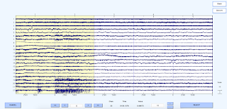
With only a week of immersion left, I have been focusing all my time on developing a preliminary proof-of-concept protocol for automating EEG artifact removal in MATLAB. Admittedly, it has been very challenging at times, especially given my limited neurology and programming experience, but I have made significant progress as of recently with the assistance of Dr. Mangat and Dr. Forgács. Last week, I began using EEGLAB, an interactive EEG analysis toolbox in MATLAB, to perform independent component analysis (ICA) on selected datasets from the subarachnoid hemorrhage (SAH) study. After ICA is applied, EEGLAB has different built-in signals processing functions to mark and remove components containing any artifacts. With several methods to choose from, I consulted with Dr. Forgács on Thursday to seek his clinical expertise. Figure 1 . Individual component analysis (ICA) to reject artifacts by visual inspection of continuous EEG signals  Figure 2. Removing ICA components using

Week 6: Regan

The vicissitudes of this week were personally testing. I had a major upset with my research when we ran out of our endothelial cell media since it has been backordered from Lonza for weeks now. Without media to feed my cells and keep them alive, I was unable to perform any experiments this week. Instead I turned to focus more on developing my clinical experiences in hematology-oncology. Firstly, I had the serendipitous opportunity to attend the final Grand Rounds presentation in heme/onc entitled "Genomic Advances in CLL" given by Dr. Andrew Lipsky, MD. The basis of the talk was to outline the current understanding of the rational for treatment order in chronic lymphocytic leukemia. Lipsky covered that CLL is a hematological malignancy found in elderly patients with a lot of variability in the clinical course required. From his talk I learned that all CLLs are cancers of B cells that occur after the B cells encounter antigen. Malignancies in this stage are thought to occur

NYC Chronicles (Week 6): More Seminal Vesicles and Stephen Colbert

Image
This week during my summer immersion experience, I took a break from my weekly visits to Columbia University Medical Center and decreased my hours of shadowing Dr. Prince in order to spend more time collecting and analyzing data for my project. In addition to compiling data, Dr. Prince and I began writing a research paper on my project regarding the dilation of seminal vesicle lumen diameter in patients with autosomal dominant polycystic kidney disease (ADPKD). The goal of my project is to investigate the genetic disease of ADPKD patients with and without dilated seminal vesicles and to correlate this information with the severity of their renal and extrarenal cystic conditions. (Figure 1, Reis et al ) Thus far, we have noticed that ADPKD patients will have developed either dilated or normal seminal vesicles (SV) after puberty (Figure 2), which remains constant over time, and that patients with a specific type of PKD mutation will be more susceptible to developing enlarged seminal ves

Week 6: Almost at the End

Image
Guy Scuderi Wow, I cannot believe week six has already come and gone. This experience has been quite remarkable so far. On Monday, I attended several meetings in the cardiothoracic surgery department where the cardiothoracic fellows presented various cases they were faced with over the past week. Although I was only able to understand about half of the terminology that was said during the presentations, it was still very interesting to absorb all this information and learn about these surgeons everyday lives and the substantial complicated cases they are faced with every day of the year. On Tuesday, I went to a three hour didactic presentation on aortic valve pathogenesis and treatment/replacement techniques presented by another cardiothoracic fellow. This was again a very interesting experience as the surgeons went over the variety of procedures that they perform to treat aortic valve diseases, specifically aortic valve calcification. Aortic valve calcification is a degenerative dis

Week 6: Carolyn

Image
This week was filled with clinical exposure, and I was surprised to see even more types of patients. As I see more and more of these surgeries, it is clear that even a specialist such as Dr. Bostrom is constantly presented with challenges throughout his career. Patrick and I were able to shadow together on Monday and observe two interesting knee cases. One case involved re-aligning the patellar implant to improve the comfort and abilities of the patient. This was particularly interesting because it involved altering the soft tissue around the knee. The procedure still required a large incision, however the surgical site did not need to be nearly as large as the primary replacement surgical site. Much of the soft tissue remained in place throughout the procedure. We also observed a conversion procedure from a unicompartmental knee replacement (prosthesis on only one femoral condyle) to a full knee replacement (prosthesis spanning the distal femur and proximal tibia). Later in t

Week 5 (Moni)

Image
This was a productive week for me, in that I finally had enough cells from my biceps tendon surgical discards to collect some flow cytometry data! The majority of the week leading up to the experiment, I spent a lot of time optimizing antibody concentrations, planning, and reading experimental protocols. The technicians at WCMC's core ran my samples for me, but I need to get trained on the analysis software to look at trends in my data. I'm hoping that next week I can analyze this round of data, and also run some more of my samples. I've also spent the week helping with some histology analysis of tendinopathy, and practicing surgical procedures on mouse cadavers. I spent the past weekend exploring much of the city, and went to the Met, Little Italy, China Town, the Brooklyn Bridge, and the Village. I also went to the American Museum of Natural History, where they have a special Mummy exhibit right now. This was really cool, because they took CT images of the tombs a

Week 5: Jeremy Keys

My fifth week has been rather uneventful as I have been getting started on the project that I was first assigned at the end of last week.  Regarding my workload, I have transitioned from doing clinical work and observing surgeries over the course of the week to primarily focusing on the analysis of data that had been previously collected from patient samples.  Unfortunately, I have been struggling a bit to interpret the existing data, as well as struggling to find an “in” to interpret the data in a meaningful way.  I am certainly interested in working through the results though, as the work pertains to how the experimental trial using copper depletion to prevent cancer recurrence in advanced stage breast cancer patients, and this may have salient ties to my research project at Cornell studying the mechanisms of cancer cell migration to form metastases.  Ultimately, now that I have gotten through the majority of primary literature relating to the copper-related proteome, and the influe

Week 5

Image
Another week of office hours, surgeries, and lab work, another week of being immersed. This week I was able to observe a case that directly utilized engineering and mechanobiology concepts. Surgeries generate a wound whenever incisions are made, making wound closure a procedure that is common to almost all surgeries. It is important that the incision is closed properly so it can heal and aid in recovery. Plastic and reconstructive surgeons place emphasis on this part of the surgery to minimize scarring and preserve optimal function. Some procedures complicate or prevent wound closure, such as when a large void in tissue is created. In the case I observed, much of the tissue was necrotic and had to be debrided. Because of this, there wasn’t enough tissue left to suture and cover the wound. One common remedy to this issue is the use of skin grafts, in which tissue from another part of the body is grafted onto the defect site. However, this creates another wound at the donor site, and th

Week5

Image
For my project, I did a summery of what I've done and made comparison between different algorithms I have tried for this project. I also read another paper of an interesting algorithm. Apart from my project, I also had a chance to go to OR and shadowed two surgeries, although I didn't make it to the end because I wasn't feeling well that day. Fig 1a: Neuron outlines detected by watershed algorithm; Fig 1b: Neuron outlines detected by region-growing based method.  Project updates: By far, I have tried three methods for automatic image segmentation: watershed, watershed-based neutrosophic method, and regrowing-based method. The first method, watershed, works very well for most of the neurons, but neurons in the region of low signal cannot be detected. A neutrosophic methods is developed based on watershed. In this algorithm, data is converted from image domain to a neutrosophic domain. However, several non-linear transformations amplify the noise, and therefore de

Week 5: MRI and Tendinopathy

Image
This week we performed additional MRI scans, this time on a whole rat sent from the Andarawis-Puri Lab in Ithaca. Our lab uses a fatigue loading procedure on rat patellar tendon as a model for overuse tendinopathy. This involves clamping the patella and tibia and applying cyclical loads that induce sub-rupture fatigue damage. This model has been used to study changes in gene expression, tissue structure, and mechanical properties and has provided insight into mechanisms of disease and healing. MR imaging, including T2* quantification, could be a useful tool to determine changes in collagen alignment and water content in the patellar tendon in vivo  after fatigue loading. This could lead MRI based techniques to quantitatively and non-invasively assess or diagnose tendinopathy. One of the challenges in scanning rats is their relatively small size when compared with humans. We attempted to use a wrist coil and a custom made rat coil to improve image quality. In addition, positioning of

Week 5: 3D bioprinter

Image
Except for shadowing Dr. John Kennedy, I have been working on the project which is to create a replacement for ankle injuries. Since nothing was set up, I had to figure it out from bottom, such as what kind of bioprinter, biomaterials, and the structure of the scaffold. We have come to a conclusion to buy a bioprinter from CELLINK, and I pushed myself to learn several programs required for designing 3D models. There are a lot of programs used for the 3D modeling; however, considering cost and UI, I have decided to use Autodesk Fusion 360. This program is super straightforward that I was able to design a simple thing after watching several tutorials. Briefly, the aim of the project is to develop the scaffold containing three layers, which are corresponding to trabecular bone, subchondral bone, and articular cartilage. The primary design of the scaffold below does not look good, but I am pretty happy with that I have reached to a level to design something. I know that there are still

Week 5: Loaded in the City

Image
Tibra Wheeler My weekend away from the city was relaxing, but I was ready to come back to the fast life. This week was filled with much time spent in lab. Monday morning I was traveling back to NYC, but that afternoon I met with Tony to watch him and another student strip some cadaver knees. The knees must be stripped of the tissue and muscle so that they can be potted for the knee simulator. There were two knees that needed to be stripped that day. We really wanted to make sure that the knees that will be used on the knee simulator are healthy knees. This was the case for the first knee that was stripped. There was lots of synovial fluid in the joint space, the bone was smooth, and the cartilage was healthy (left picture). Unfortunately, this was not the case for the second knee. The second knee was very inflamed, there was blood in the synovial fluid, and much of the cartilage had become mineralized and was hard (right picture). It was really interesting to see this because